PLATE XII. [PLATE 311].
[395] ALTHOUGH the scorpion has been made the subject of repeated investigations by some of the best minute anatomists of past and present times, it is a remarkable circumstance that no exact account of the structure of the commencement of its alimentary canal is to be met with, at least so far as my knowledge extends. Meckel ('Beiträge zur Vergleichenden Anatomie,' Band 1, Heft 2, 1809), as might be expected from the fact that his dissections were performed without the aid of even a magnifier (page 106), takes no particular notice of the small and delicate parts in question. Treviranus ('Bau der Arachniden,' 1812) is equally silent as to this important portion of the economy of the scorpion; and even the accurate Johannes Müller, in the essay entitled "Beiträge zur Anatomie des Scorpions" (Meckel's 'Archiv.,' 1828), which threw so much new light upon the organization of this animal, although he saw more than either his predecessors or his successors have done, did not probe the matter to the bottom. In describing the alimentary canal, he merely says:–"The pharynx which arises in front of the brain, upon a particular, strongly excavated, portion of the skeleton, is much wider than the rest of the intestine, and resembles a vesicle. The oesophagus is very delicate where it proceeds from this vesicle, rises between the very stout nerves for the chelæ, above the brain (which lies behind the pharynx), and passes over the saddle-shaped upper excavation of the internal thoracic skeleton, whilst the spinal cord and the posterior cerebral nerves pass through the opening of the .same skeleton."
[396] Even the elaborate and beautifully illustrated memoir on the organization of Scorpio occitanus, published by M. Blanchard, a couple of years ago,1 does not furnish the inquirer with either definite or accurate information on this point. At page 19, I find under the head of "mouth":
"In the scorpion there exists only a single buccal piece properly so called; it is inserted in the median line above (au-dessus) the mouth, just below the cheliceræ (antennes pinces), and wedged in, so to .speak, between the foot-jaws. It is a little flexible appendage, thinner towards its extremity, sensibly dilated laterally, convex above, and beset, chiefly at the end, with fine and silky hairs. This piece presents two apodemata (apodèmes d'insertion), which diverge greatly from one another.
"One finds a certain difficulty in positively determining the nature of the single buccal appendage of the scorpion. It is impossible to regard it as the analogue of the labrum (lèvre supérieure) of insects. The labrum is one of those pieces which abort most completely in the arachnida. Besides, in all articulata, this labrum receives nerves which arise from the cerebral ganglia. It is different with the buccal appendage of the scorpion; its nerves arise from the anterior part of the suboesophageal ganglia, exactly like those of the mandibles and maxillæ of Crustacea and Insects. It can thus only be compared to these pieces; but ought we to regard it as representing both the mandibles and the jaws, or only the mandibles, or the jaws, either one or the other being supposed to be aborted?"
With respect to both the main points contained in these paragraphs, however, M. Blanchard subsequently makes statements which seem difficult to harmonise with the conclusions enunciated.
Thus, at page 41, I find:
"The pharyngeal nerves are two pair. Those of the first take their origin from the anterior and median edge of the cerebrum, and almost immediately unite so as to form a single nerve, whose branches are distributed in the upper portion of the buccal appendage. It is evidently the analogue of the nerves of the labrum,of insects."
And, again, at page 60
" Mouth and oesophagus.–The buccal orifice appears under the form of a little transverse cleft, hidden under the cheliceræ above (au-dessus) the median appendage, which has already been described (p. 19); its edges are flexible, and are deprived of asperities. The oesophagus, which commences in a slightly funnel-shaped pharynx, is [397] delicate, short, and widened posteriorly, so as to resemble what M. Léon Dufour calls the 'jabot' in insects. The oesophagus is held upon each side, towards its middle, by a fine muscular band directed backwards, and towards its point of union with the stomach by a similar band directed forwards. These muscles are attached to the sternal floor, formed, as is known, by the basilar pieces of the appendages. They serve to stretch the oesophagus either forwards or backwards, so as to facilitate deglutition.
"The walls of the oesophagus are thin and smooth internally, and present a few fine folds."
In the figures (op. cit., pl. iv, figs. 1 and 6), which represent the anterior part of the alimentary canal, the oesophagus is represented as a straight, taper tube, ending in the mouth, without change of direction.
At page 32, M. Blanchard states, under the head of–
"Muscles of the buccal appendage.–We have indicated the two, long,. diverging, apodemes of this piece (p. 19). Upon the base of each of them is inserted an elevator muscle, provided with two fixed attachments to the cephalo-thoracic shield in front of and external to the median eyes (P1. ii, fig. 4 e e and fig. 6 a). By its contraction, this muscle causes the buccal appendage to be elevated a little–a movement which takes place when the animal introduces food into its mouth. A transverse muscle is attached to the two apodemic plates (P1. ii. fig. 41); it is this muscle which, acting either on the one side or on the other, determines the slight lateral movements of the buccal appendage. It is to be observed, that this piece, solidly fixed between the foot-jaws, sensibly involves the latter during the execution of its slight movements."
The structure of the parts which I have observed in a large species of Buthus may be described as follows:
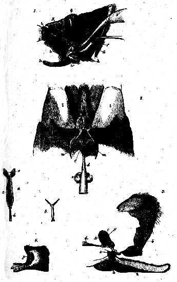
The "buccal appendage" of M. Blanchard is a vertically elongated, laterally compressed, cushion-like prominence, broad and rounded above, where it is marked by a slight median ridge, slightly concave from above downwards in front, and narrowed below (P1. XII, figs. 1, 2, 3 b). Its anterior and lateral surfaces are covered with fine, short hairs, which form a projecting pencil at its anterior inferior angle. There is no aperture whatsoever above this body, between the cheliceræ; but, below and behind it, the aperture of the mouth, large enough to admit the head of a fine needle, can be very easily found. I entertain no doubt, therefore, that this "buccal appendage" is a true labrum, and, indeed, in all essential respects, it is exactly like that part in the crustacea
[398] The convex lower surface of the labrum bounds the mouth in front, while behind, it is limited by a transverse thickening of the chitinous integument, which appears to represent the sternum of the mandibular somite (fig. 4 o). The mouth opens into a very curious pharynx, formed by a delicate outer investment, and a strong inner chitinous lining. Viewed laterally, this organ (c) has the shape of a pear, its broad end being uppermost, and its long axis directed obliquely upwards, and backwards, in such a manner, that the broad upper end lies in the middle, between the prongs of the fork-like apodeme, which M. Blanchard has described. Viewed from above or below, however, the pharynx appears to be very narrow, indeed, almost linear, in consequence of its very peculiar form, which is displayed in the section, taken transversely to the longitudinal axis and perpendicularly to the vertical plane represented in fig. 5. The cavity of the sac is here seen to be triradiate, while its walls are very closely approximated, so as to leave but a slight interspace. The narrow band which joins the two lateral walls below and behind is slightly excavated, so as to present a convexity towards the cavity of the pharynx. The two shorter rays of the sac are turned upwards and outwards; the third longer ray is directed vertically downwards. The oesophagus, an exceedingly delicate and narrow tube, comes off from the posterior wall of the vertical ray or crus of the pharynx, just above the mouth; and, widening, passes backwards and upwards, into the dilatation which receives the ducts of the so-called salivary glands (e). just above the aperture is a rounded projection (fig. 6 p), which I suspect may act as a sort of valve, when the sides of the pharynx are divaricated, by more or less completely occluding the oesophageal aperture. The inner surface of the chitinous lining of the pharynx is more or less rugose: and, towards the oesophageal aperture, presents a number of very minute spines (fig. 6).
The transverse muscular fibres (fig. 2 n), rightly said by M. Blanchard to arise from the forks of the apodeme (m), are inserted into the side walls of the pharyngeal sac, which is so narrow from side to side, as readily to escape notice, without dissection. The termination of the aorta appeared to me to pass between the two superior crura of the sac.
The large vertical muscles (fig. 1 q) are, as M. Blanchard states, inserted into the base of the apodeme; and, besides these, the labrum is traversed by strong transverse and longitudinal muscles.
The mode of action of this curious apparatus appears to be readily intelligible. Scorpions, as is well known, suck the juices of their prey, and the pharyngeal sac seems to be well calculated to [399] perform the part of a kind of syringe. For, suppose the prey to be held between the labrum above, the bases of the great mandibles of the sides, and the processes furnished by the maxilliary limbs below, and that the minute oral aperture is applied to a wound. Then, if the transverse muscles (n) contract, the sides of the pharynx will be drawn apart, and a partial vacuum, or at least a tendency to the formation of one, will be created. If, by the same action, the projection (p) is brought down over the oesophageal aperture, regurgitation from the oesophagus will be prevented; but, in any case, as the oral aperture is larger than the oesophageal, it will be easier for the sac to be filled through the mouth. The sac being full, if the labrum is depressed so as to close the oral aperture, and the transverse muscles are relaxed, the elasticity of the walls of the pharynx will tend to reduce its cavity to its primitive dimensions, and hence to drive the ingested liquid into the oesophagus. Successive repetitions of the action would gradually pump the juices of the prey into the alimentary canal of its captor.

Fig.
1.–Longitudinal vertical section of the cephalo-thorax of a Scorpion, showing the pharynx, oesophagus, nervous centres, and the large eyes in their natural relations.
2.–Dorsal view of the cephalo-thorax of a Scorpion. opened and dissected, so as to show the apodemata, and the anterior portion of the alimentary canal, with the pharyngeal muscles.
3.–The chitinous lining of the anterior part of the alimentary canal, the integument of the labrum. and the basal processes of the first maxilla.
4.–The chitinous lining of the pharyngeal sac, viewed from above.
5.–A transverse section of the same, taken along the line x y (fig. 3).
6.–The region of the pharyngeal sac near the commencement of the oesophagus.
The letters have the saire significations throughout::–a, mouth; b, labrum; c, pharynx; d, oesophagus; e, salivary duct; f, diaphragm; g, eye and ocular nerve; h, suboesophageal ganglion; i, antenna; k, maxilla; 1, mandible; m, apodeme; n, pharyngeal muscles; o, sub-oral transverse thickening of the chitinous integument; p, valve (?) of the pharynx; x y, line along which the section in fig. 5 is taken.
1 The livraisons of M. Blanchard's work are unfortunately published without dates.
|
THE
HUXLEY
FILE

|
| ||||||||||||||||||||||||||||||||||||||||||||||||||||||