Medallion
by Frank BowcherHuxley Memorial Committeee (1889)
Scientific Memoirs IV Frontispiece
[26] THE singular little fish Amphioxus lanceolatus has been universally regarded as an extremely anomalous member of the Vertebrate series by reason of the supposed absence of renal organs and of any proper skull and brain. On these grounds, chiefly, Agassiz proposed to separate it from all other fishes, and Haeckel, going further, made a distinct division of the Vertebrata (Acrania) for its reception; while Semper,1 in a lately published paper, separates it from the Vertebrata altogether.
In a recent communication to the Linnean Society I have described what I believe to be the representative of the ducts of the Wolffian bodies, or "primordial kidneys" of the higher Vertebrata, in Amphioxus; and I propose, in this preliminary notice, to point out that although Amphioxus has no completely differentiated brain or skull, yet it possesses very well marked and relatively large divisions of. the cerebro-spinal nervous axis and of the spinal column, which answer to the encephalon and the cranium of the higher Vertebrata.
The oral aperture of Amphioxus is large, of a long oval shape, and fringed by tentacles, external to which lies a lip, which is continuous behind with the ventro-lateral ridge of the body. The oral chamber is spacious, and extends back to the level of the junction between the sixth and seventh myotomes (fig. A). Here it is divided from this branchial cavity by a peculiarly constructed muscular velum palati, the upper attachment of which to the ventral aspect of the sheath [27] of the notochord lies vertically below the anterior angle of the seventh myotome.
Eight pairs of nerves are given off from the cerebro-spinal axis as far as this point. The eighth, or most posterior of these, which, for convenience, may be called h, passes out between the sixth and seventh myotomes, and runs down parallel with the lateral attachment of the velum. The next five (g, f, e, d, c) pass out between the first six myotomes, and are distributed by their dorsal and ventral branches to those myotomes, to the integument and to the walls of the buccal cavity. The foremost two nerves (b and a) pass in front of the first myotome, and the nerve a runs parallel with the upper side of the notochord to the end of the snout, giving off branches to that region of the body which lies in front of the mouth. This nerve lies above the eye-spot.
In the Marsipobranch fishes Myrine and Ammocætes (now known to be a young condition of Petronzyzon) a velum also separates the buccal from the branchial cavity (figs. B, C, D). But this velum is in connexion with the hyoidean arch. The resemblance of the buccal cavity, with its tentacles, in Ammocætes to the corresponding cavity in Amphioxus is so close that there can be no doubt that the two are homologous. In the Ammocætes there is a hyoidean cleft which has hitherto been overlooked. The auditory sac lies at the dorsal end of the arch and above the dorsal attachment of the velum. The latter, therefore, corresponds with the auditory region of the skull, and the nerve h should answer to the last of the præeauditory cranial nerves, which is the portio dura. Assuming this to be the case, though the detailed homologies of the cranial nerves of the higher Vertebrata are yet to be worked out, it follows that the segment of the cerebro-spinal axis which in Amphioxus lies between the origin of the nerve h and the eye, answers to all that part of the brain which lies between the origin of the seventh nerve of Petromyzon and the optic nerve. Consequently, the lateral walls of the neural canal in the same region answer to that region of the, skull in Petromyzon which lies between the origin of the seventh and the origin of the optic nerve. Hence, as each myotome of Amphioxus represents the corresponding portion of a protovertebra, it follows that the same region of the skull in the Lamprey and other Vertebrata represents, at fewest, six protovertebræ, almost all traces of which are lost, even in the embryo condition of the higher Vertebrata.
It may further be concluded that the several pairs of nerves which leave the cerebro-spinal axis, between those which answer to the poitico dura and the optic nerve, in Amphioxus, are represented by the third, [29] fourth, fifth, and sixth pairs of cranial nerves of the higher Vertebrata. The nerve a, in fact, has the characteristic course and distribution of the orbito-nasal division of the trigeminal; while, without at present drawing a closer parallel, it is easy to see that the nerves b, c, d, e,f, and g., with their respective myotomes, supply the requisite materials for metamorphosis into the oculomotor, pathetic, trigeminal, and abducens nerves, with the muscles of the eye and of the jaws, in the more differentiated vertebrate types..
Thus, that part of the cerebro-spinal axis of Amphioxus which lies in front of the seventh myotome answers to the præauditory part of the brain in the higher Vertebrata, and the corresponding part of the head to the trabecular region of the skull in them. On the other hand, from the seventh myotome backwards, a certain number of segments answer to the post auditory, or parachordal region of the skull of the higher Vertebrata. .
The answer to the question, how many? involves sundry considerations. It must be recollected that though the branchial chamber of Amphioxus is the homologue of the branchial chamber of other Vertebrata, it does not necessarily follow that the imperfect branchial skeleton of Amphioxus corresponds with their branchial skeleton. The branchial skeleton of the higher Vertebrata consists of cartilaginous rods, which seem to be developed in the somatopleure, and to be homologous with the ribs, while the branchial skeleton of Amphioxus consists of fibrous bands apparently developed in the splanchnopleure.
The branchial arches of the higher Vertebrata, in accordance with their essentially costal nature, receive their innervation from the glossopharyngeal and pneumogastric nerves, which are homologues of spinal nerves; and, in seeking for the posterior limits of that region in Amphioxus which corresponds with the skull and brain in other Vertebrates, we must only take into account as many pairs of those nerves which arise from the cerebro-spinal axis as we know are, in the Vertebrata next above Amphioxus, devoted to the branchial arches. In none of these are there more than seven pairs of branchial arches; so that not more than eight myotomes (and consequently protovertebræ) of Amphioxus, in addition to those already mentioned, can be reckoned as the equivalents of the parachordal region of the skull in the higher Vertebrates. Thus it would appear that the cranium of the latter is represented by those segments of the body of Amphioxus which lie in front of the fifteenth, counting from before backwards, and that their cranial nerves are represented by the corresponding anterior pairs of nerves in Amphioxus.
[30] In all Vertebrata above Amphioxus the nerves which answer to the seven posterior pairs in Amphioxus unite into one or two trunks on each side, and give rise to the nerves called pneumo-gastric and glosso-pharyngeal; and, as these pass out of the skull in front of the occipital segment, it would appear that this segment is, in the main, the result of the chondrification, with or without subsequent ossification, of the fourteenth protovertebra.
There is no evidence, at present, that the ear-capsule represents a modification of any part of the vertebral skeleton, nor that the trabeculæ are anything but an anterior pair of visceral arches. And if these parts have nothing to do with centra, or arches, of vertebræ, it follows that the numerous protovertebræ, which lie in front of the fourteenth in Amphioxus, are represented only by muscles and nerves in the higher Vertebrata.
The anterior end of the cerebro-spinal axis of Amphioxus answers to the lamina terminalis of the thalamencephalon of the higher Vertebrata, the cerebral hemispheres and olfactory lobes remaining undeveloped.
If the auditory nerve is, as Gegenbaur has suggested, the dorsal branch of a single nerve which represents both the portio dura and the portio mollis, the auditory organ of Amphioxus is to be sought in connexion with the dorsal branch of its eighth nerve. I have found nothing representing an auditory organ in this position; and I can only conclude that Amphioxus really has no auditory apparatus. In all other respects, however, it conforms to the Vertebrate type; and considering its resemblance to the early stages of Petroinyzon described by Schulze, I can see no reason for removing it from the class Pisces. But its permanently segmented skull and its many other peculiarities suggest that it should be regarded as the type of a primary division or subclass of the class Pisces, to which the name of Entomocrania may be applied, in contrast to the rest, in which the primary segmentation of the skull is lost, and which may be termed Holocrania. On a future occasion I propose to show in what manner the skull of the Marsipobranch is related to that of the higher Vertebrata, and more especially to the skull of the Frog in its young tadpole state.
[28]
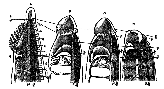
A, C, D are diagrammatic, but accurate, representations of the anterior part of the body in Amphioxus (A), in an Amnocæte 1.6 inch long (C), and in a fully grown Ammocoete 5.7 inches long (D). B is a copy of the furthest advanced stage of the young Petromyzon planeri six weeks after hatching, as figured by Schultze in his memoir on the development of that fish. The figures are magnified to the same vertical dimension, so as to afford a means of estimating, roughly, the changes in the proportional growth of the [31] various parts of the head of the Lamprey in its progress from the embryonic towards the adult condition. In C, the brain is already differentiated into the three primary vesicles and the vesicles of the cerebral hemispheres, though they are not shown, the whole brain being merely indicated by the dark shading. The trabeculæ (Tr), which have already united in front, are indicated, but not the semilunar ethmoidal cartilage, which lies above and behind the nasal sac. In D, neither the ethmoidal nor the trabecular cartilages are shown, but the contour of the brain is indicated; and the manner in which the longitudinal muscles, which represent the anterior myotomes of Amphioxus, are arranged is shown. The tentacles of Amphioxus are represented by the tentacles of the Ammocoerte, the hood-like "upper lip" of the latter obviously answering to the median prolongation of the head of Amphioxus with the two lateral folds of integument which lie outside the bases of the tentacles and are continued back into the ventrolateral ridges. The relative shortening of the notochord, and lengthening of that region of the brain which lies in front of the origins of the optic nerves, in C, as. compared with B, is remarkable.
A line is drawn in all the figures through the anterior margin of the nasal sacs. (Na-Na); another has the same relation to the eyes (Op-0p); and a third (Hy-Hy) passes through the region of the auditory sac and hyoidean arch. 1, 2, 3, hyoidean and first and second branchial clefts of Ammocoetes; i., ii., iii., iv., &c., myotomes of Amphioxus; My, myelon or spinal cord; Ch, notochord.
1 "Die Stammverwandtschaft der Wirbelthiere und Wirbellosn," Arbeiten aus dem zool.-zootoni. Institut in Wiirzburg, Bd. ii. 1874, P. 42.
|
THE
HUXLEY
FILE
|
| ||||||||||||||||||||||||||||||||||||||||||||||||||||||